Information that goes into your brain can lead to two potential
actions: thought and movement. Because Testosterone is all about movement, I'm going to outline how your brain leads to
muscle contraction. I'm also going to discuss fatigue, and tell you
what structures are key to regulating your movements. Put on a pot
of coffee and take the phone off the hook because this ain't
exactly light reading.
Communication in the nervous system is electrochemical. That's
just like it sounds: a combination of electrical and chemical
activity. For one nerve to communicate with the next, an electrical
signal is sent down the nerve's axon to the post-synaptic terminal.
This electrical activity at the end of the terminal results in
chemical changes that dump neurotransmitters out into the synapse.
The neurotransmitter attaches to receptors on the post-synaptic
cell and causes that cell to activate (depolarize).
 |
Neural activity: an electrochemical
orgy.
Now let's examine how your biceps contracts. Every conscious
movement starts in your brain. Your brain sends a descending signal
down your spinal cord to the motor neuron that exits to your
biceps. In this case we're talking about the cervical vertebrae 5
and 6 (C5 and C6). The motor neuron exits your spinal cord at C5-C6
and then merges into your biceps. Where the motor neuron meets your
biceps is known as the motor end plate. This is the space
between the motor neuron and your muscles.
 |
Typical motor unit.
The electrical activity in the motor neuron, from your brain,
reaches the motor end plate, triggering the release of the
neurotransmitter acetylcholine. Acetylcholine attaches to
acetylcholine receptors in the muscle, allowing sodium to rush in
and potassium to rush out. This is known as depolarization.
This depolarizing causes a release of calcium in your muscles.
The calcium binds to the actin filament and allows it to interact
with myosin. When myosin and actin attach, your muscles contract.
ATP fuels the contractions by allowing actin and myosin to unbind
so they can reattach and contract further. If ATP isn't present,
actin and myosin can't unbind and your muscles end up in rigor
mortis. As long as ATP and calcium are present, the muscles can
continue to contract.
Muscle Fibers and Fatigue
When muscle fibers drop out due to fatigue, it's because there's
not enough ATP to fuel the contractions. Here's a basic overview of
how the big, medium, and small muscle fibers fatigue with
contractions.
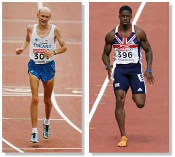
Big Muscle Fibers (type IIB/X) - Your largest muscle fibers rely
on the ATP-PC energy system that can't generate much ATP. The
ATP-PC system can only produce ATP for 15 seconds, max. In most
cases, the time before depletion is closer to 8 or 10 seconds.
That's why your biggest muscle fibers drop out first, and why you
can't sustain maximum voluntary contractions for more than 8-15
seconds.
Medium Muscle Fibers (type IIA) - Your medium sized fibers rely
on anaerobic glycolysis, which is an energy system that can produce
more ATP than the ATP-PC system. That's why your medium sized
fibers can contract for longer periods of time. Anaerobic
glycolysis can produce contractions on the order of minutes, with
10 minutes probably being the upper end.
Small Muscle Fibers (type I) – The smallest muscle fibers
can maintain contractions for hours at a time because they rely on
aerobic metabolism to produce ATP. Of course, "aerobic" means "with
oxygen." Since there's usually plenty of oxygen, there's also
plenty of ATP.
 |
Small muscle fibers (left) vs. big muscle fibers (right).
Got it? Let's get back to the motor end plate.
Bridging the Gap
When acetylcholine is released at the motor endplate, it
attaches to specific receptors on the muscle that leads to the
steps I outlined earlier. But what happens to the acetylcholine
after you've stopped flexing your arm? After all, if acetylcholine
hung around your muscles wouldn't stop contracting. In other words,
acetylcholine needs to be inactivated.
After you're finished flexing your biceps for the busty fitness
babes, acetylcholine is broken into pieces (inactivated), then
transported back up to the pre-synaptic cell to be re-synthesized
so it can be released again. This is a very efficient process since
it's localized. A neurotransmitter needs to be taken up directly at
the site of release so your nerves can react very quickly to the
nervous system's demands.
On a similar note, the drug Prozac directly affects this
re-uptake process, but for a different neurotransmitter, serotonin.
This leaves you with more serotonin floating around to keep
activating the post-synaptic cell. For many people, this augments
their sense of well-being (hence the term anti-depressant).
Interestingly, cocaine affects the same mechanism as Prozac, along
with many others that make it, well, not so good for you.
What happens if your nerves can't release acetylcholine? You get
paralysis. This is how neurotoxins work. A neurotoxin is a
substance that negatively affects nerve transmission. Sounds kind
of nasty, doesn't it? Well, middle-aged women, metrosexuals, and
Joan Rivers simply adore the paralyzing effects of the
neurotoxin botulinum toxin.
Don't let the innocuous name fool ya: it's actually the most
toxic protein in the universe. In case you hadn't guessed, it's
that crazy little chemical that delights doctors and mortifies
neuroscientists: Botox.
 |
Can we talk about deadly paralyzing
neurotoxins?
I'm spending some extra time discussing acetylcholine because
many fly-by-night supplement companies have attempted to sell you
on the idea that they can increase the release of acetylcholine at
the motor end plate. More acetylcholine means more muscle, right?
Wrong. There's plenty of acetylcholine to go around. That's why
your body has mechanisms to inactivate it and transport it back
into your nerves.
I started this discussion by skipping over your brain. I've
found it's easier for people to learn new information at the muscle
level first. Once they have a handle on that, it's usually more
effective to move upward – to the brain – where things
can get a wee bit more complicated.
Back to the Brain
The cortex is the outer most area of your brain. It's a pale
gray structure, and looks kind of like a mushroom with all of its
peaks and valleys (these are known as fissures). If you flattened
the cortex out, it would be as thick as six playing cards, and the
size of a dinner napkin. The cortex is the "newest" part of the
human brain, and it's what separates us from lower species. In
other words, because your cortex is much larger than that of most
other animals, you're well-equipped to outsmart that darn cat
that's always hiding behind the curtain.
There's a right and left side to your cortex. The right cortex
controls the left side of your body; the left cortex controls the
right side. This contralateral control occurs because the nerves
cross in the lower portion of your medulla before entering the
spinal cord.
There's an anatomical representation of your entire body in a
section of the cortex called the motor cortex (also known as Area
4). If you were unfortunate enough to stand underneath a guillotine
and your ex dropped the blade so it split your cranium right in the
middle, it would slice through your motor cortex.
 |
The primary motor cortex (blue), sliced with transparent
guillotine blade.
The area towards the midline of your brain represents your toes.
As you move away from the midline and around the cortex the
physical representation of your body merges from your toes to your
shins to your thighs, etc. until it ends at your mouth.
 |
The body map of the motor cortex.
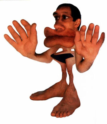
Interestingly, and not surprisingly, there's a disproportionate
representation of your body in your motor cortex. Your mouth and
fingers take up the largest percentage of the motor cortex. This
should be no surprise since each structure requires fine, complex
movements. Speaking is a complex motor process, so is playing a
Chopin tune.
 |
Your body, as represented by your brain's motor/sensory
cortex.
If I zapped your motor cortex with electricity in a specific
area, a specific set of muscles would fire. Let's say I was cut off
in traffic and I had a buddy along for the ride. If I had the
equipment, and if he was willing, I could jam an electrode into the
left side of his motor cortex at the area that represents the
middle finger. Then I'd zap a little electricity and voila!
His right middle finger would pop up. Unlikely scenario, indeed,
but it gets the point across. Here's a great link that allows you do what I just described.
So I zap the middle finger area of your motor cortex and your
middle finger ends up flying the bird. This is the direct
corticospinal pathway from your brain to your muscles. It's also
known as the descending neural tract. The corticospinal pathway is
part of the pyramidal motor system. The reason why the direct
descending pathway is called the pyramidal motor system is because
the nerves that initiate the pathway in your motor cortex are
shaped like pyramids. Hey, who doesn't love pyramids?
 |
Pyramidal neurons: everybody loves
pyramids.
You also have indirect pathways called the extrapyramidal motor
system that allows your muscles to maintain tone during everyday
activities. In other words, the muscles that allow you to remain
upright must have constant tension or else you'd fall to the floor
like a rag doll. The indirect pathway controls all of the slight
involuntary movements that you don't consciously think about, such
as when you're standing in line at the movies. But if a guy behind
you pinches your girlfriend's keister, the intense voluntary movements that follow are due to the direct pyramidal pathway.
The extrapyramidal might just be the most complicated aspect of
the motor system. It consists of many different brain areas such as
the basal ganglia and substantia nigra that are constantly
performing feedback and feedforward information that regulates the
motor cortex.
If you're having trouble differentiating between the pyramidal
and extrapyramidal motor systems, here's a tip. The pyramidal motor
system goes into the spinal cord, where it can directly affect your
muscles. The extrapyramidal motor system does not reach the spinal
cord. Instead, it adjusts and regulates the pyramidal motor system.
The motor cortex (area 4) is interconnected with sensory areas
of the cortex (areas 1-3). In other words, all four areas are
positioned next to each other in the brain. That's because your
brain requires constant feedback during any movement. As you curl a
dumbbell, information is being sent to the sensory area of your
brain, which then feeds into the motor area. This is how you
constantly adjust and regulate your movements.
How does that happen? Read on.
Sarah Bellum?
The cerebellum is an extraordinary part of your brain that sits
at the back of your head. There are approximately 100 billion
nerves in the brain, and half of them are in the cerebellum! It's
part of the extrapyramidal (indirect) motor system that's
constantly regulating and perfecting your movements.
When you watch Tiger Woods swing a club with the same precision
every time, it's because his cerebellum has produced the movement
so many times that its interaction with the descending pyramidal
motor system is smooth and efficient. On the other side of the
coin, babies that are learning to walk have a difficult time at
first because their cerebellum hasn't perfected the motor pattern.
Luckily, our cerebellum has a pretty good memory. That's why you
can ride a bike after being off one for 20 years.
How important is the cerebellum for movement? Let's say your
buddy is a tightrope walker who's also rich as hell. He bets you a
million bucks that he can walk the tight rope faster than you. You,
of course, will lose unless you can devise a devious plan. If you
smack him across the back of his head with a Louisville Slugger, it
will damage his cerebellum and he won't be able to walk, let alone
balance. (I probably don't need to mention that this information is
strictly for entertainment purposes. If you're going to smack
someone on the back of the head, make sure it's not your
friend.)
 |
Don't try this just after getting smacked in the
cerebellum.
Amazingly, the cerebellum doesn't have nerves that enter the
spinal cord. This really is incredible considering that your
cerebellum is the key to smooth, efficient movement. How does the
cerebellum affect movement? Because your corticospinal (descending)
pathway has nerves that branch off in your brain and synapse with
the cerebellum.
If you've read up on strength training you're probably familiar
with the law of repetition. It states that the more times you
perform a movement, the better you'll get. If you want to press a
lot, you've got to press a lot, they say. This is true. However,
most "experts" tell you that it's because of changes at the
neuromuscular junction. Actually, repeating a movement pattern is
fine-tuning your cerebellum.
Before I finish up I must give respect to my professors, and
mention one more thing. As with everything in the nervous system,
the pyramidal (direct) and extrapyramidal (indirect) pathways are
very closely connected, and must work together. In essence, even
the simplest movement involves hundreds of interconnected pathways.
I talk about them separately for learning purposes, but they're
forever coalesced.
Conclusion
I hope you've learned a thing or two during this journey from
your brain to your biceps. Eat your fruits and vegetables, take
fish oil, and keep lifting weights. These are the keys to keeping
your nervous system healthy.


